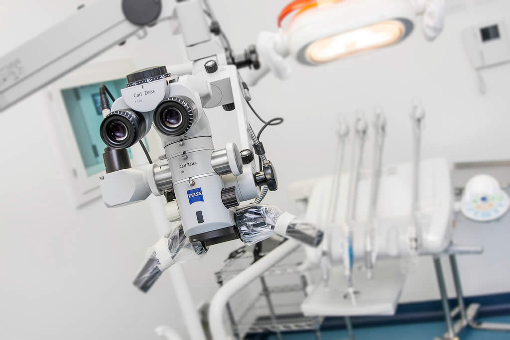
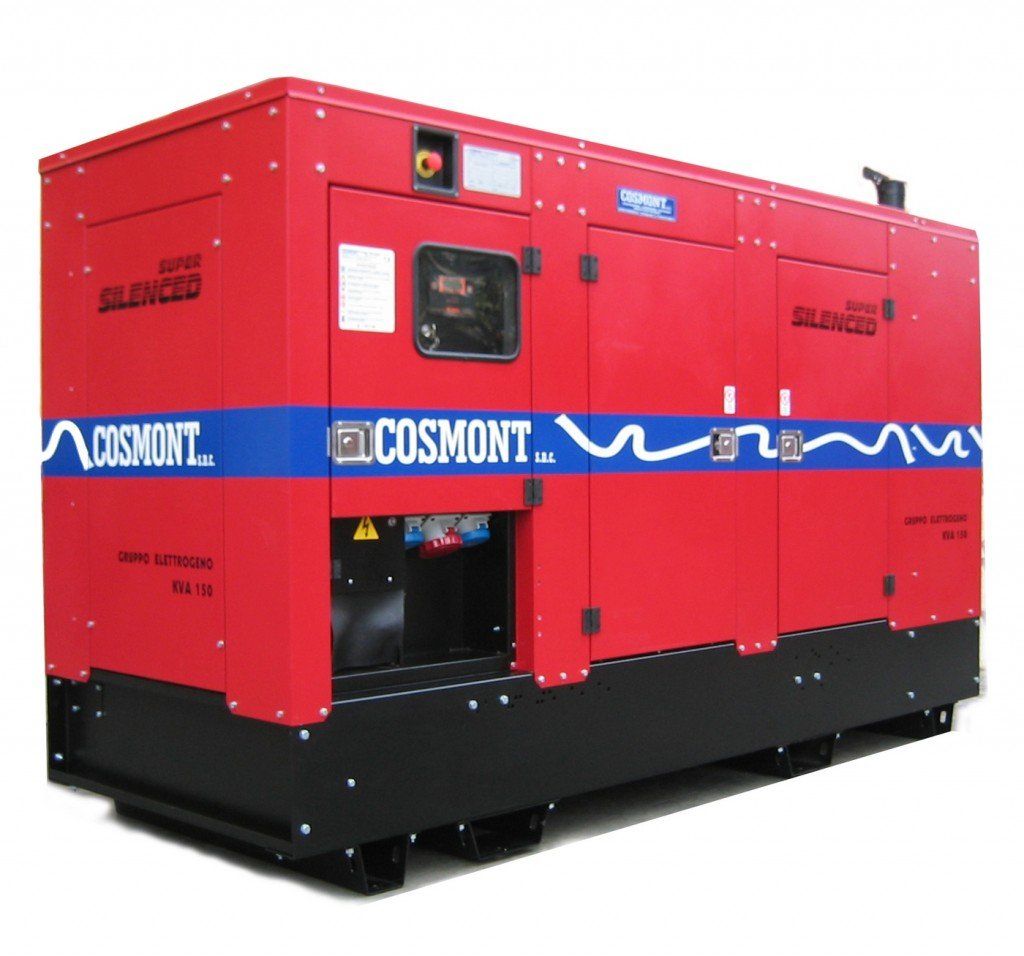
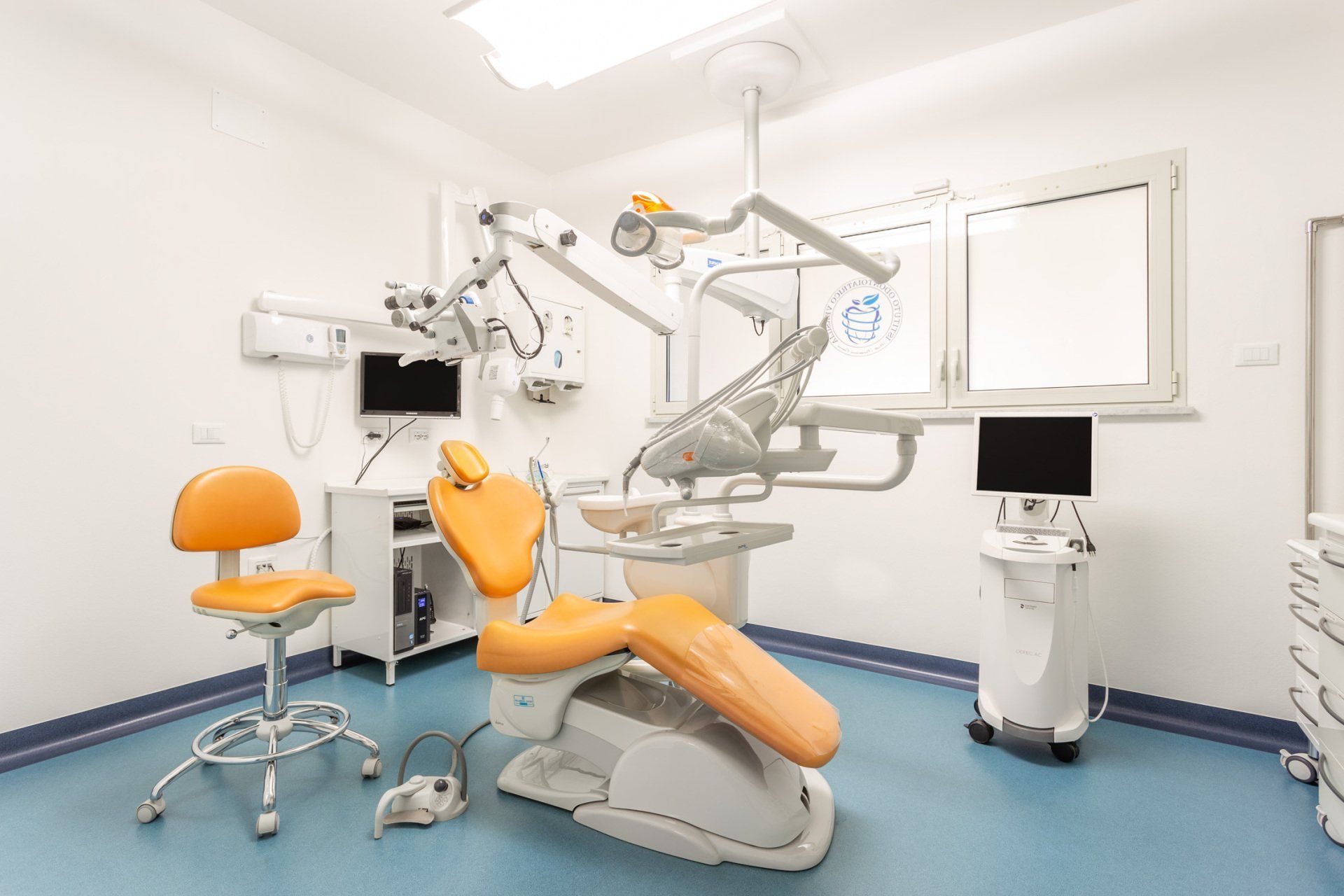
Versilia Dental Institute in Forte dei Marmi is equipped with the most modern and innovative equipment. Among these are the operating room generator and the operating microscope, which magnifies details up to 20 times, allowing the resolution of even the most complex clinical cases.
Contact the practice
Emergency and prevention
The presence of a generator in the operating room is essential for performing procedures safely: the generator at the institute in Forte dei Marmi can power operating rooms with 22kW of electricity, preventing dangerous interruptions to procedures due to potential power outages.
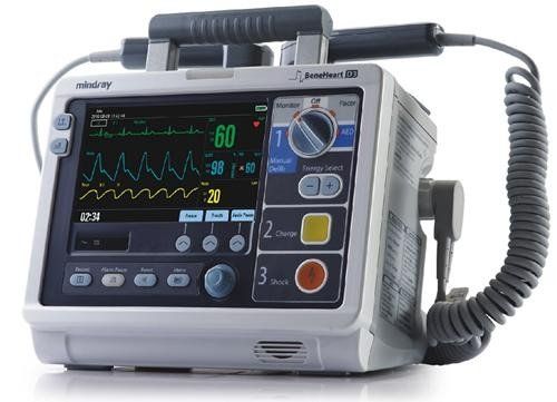
Emergency devices
At Versilia Dental Institute in Forte dei Marmi, oral and dental surgery procedures and check-ups are performed daily.
Although cardiopulmonary emergencies are rare, the practice is equipped with all the necessary technology to quickly and safely address such situations, including the operating room generator and microscope.
Conscious sedation
t
Conscious sedation
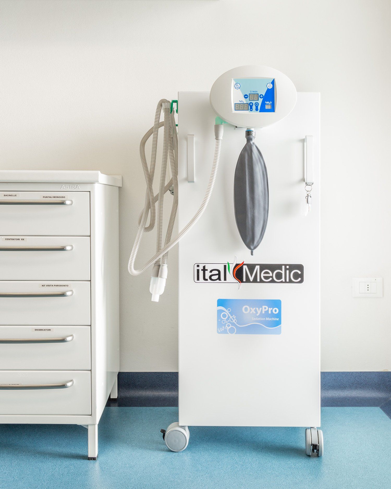
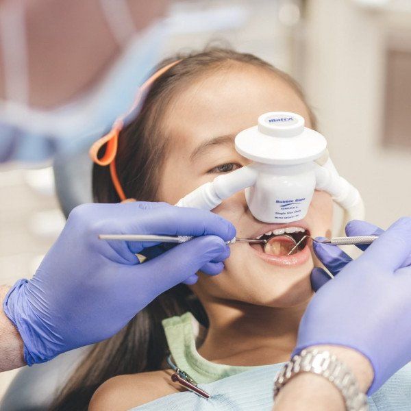
Thanks to new technologies, we can now calm our patients with nitrous oxide; inhaled aerosol
that induces relaxation and helps us manage dental appointments more calmly.
Comfort and tranquillity
Fear of the dentist causes anxiety and distress in both children undergoing dental procedures and the parents accompanying them. Eliminating these emotional states can make a visit to the dentist extremely pleasant.
For adults and children
Conscious sedation with oxygen and nitrous oxide is a sedative analgesia technique that finds its ideal application in paediatric dentistry.
What is it?
Conscious sedation (also known as sedation anaesthesia) is an anaesthetic technique with nitrous oxide and oxygen that aims to induce a state of relaxation, promoting amnesia and pain control.
The result is that the patient, while remaining conscious, feels no pain and, most importantly, has no memory of the procedure.
Radiographic technology
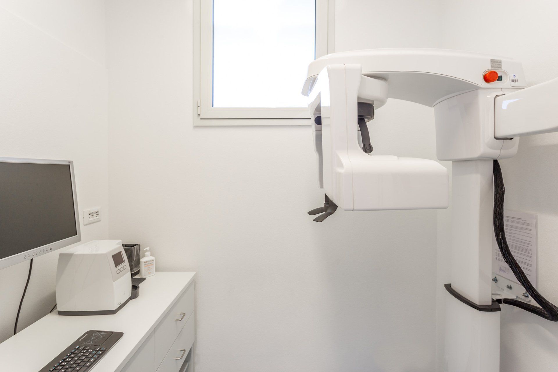
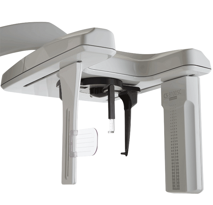
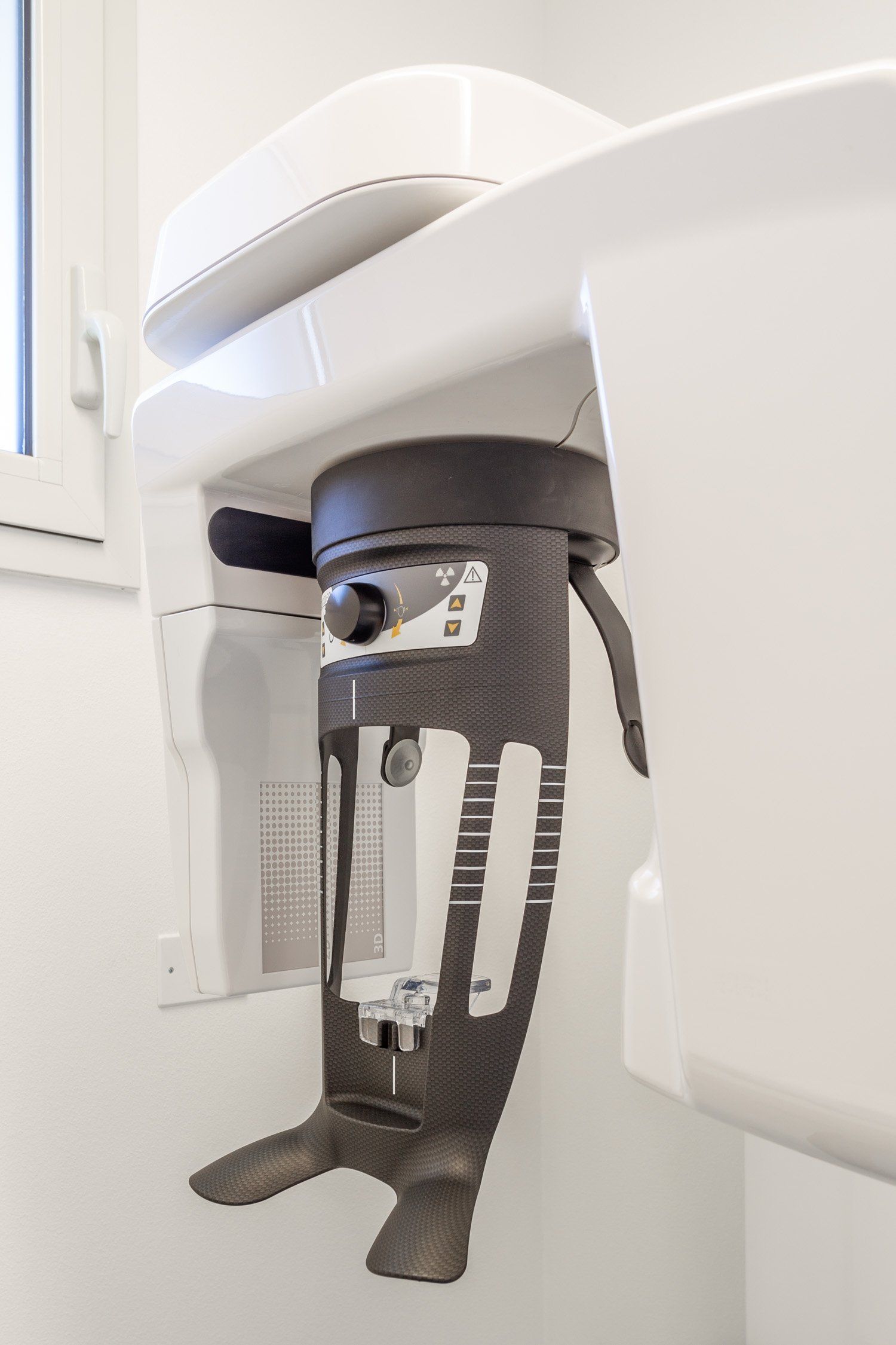
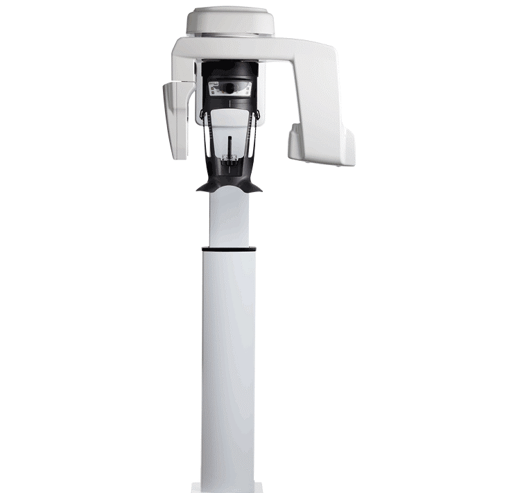
In the diagnostic phase, radiography is essential. For this reason, Versilia Dental Institute provides its patients with all the necessary technology to ensure safety during their treatments. We benefit from the presence of a Carestream radiographic machine, which allows us to acquire three types of radiographs:
- Cephalometric X-ray
- OPT (Orthopantomogram)
- Cone Beam CT
- Digital X-rays
1. Cephalometric X-ray
This method, alongside panoramic dental X-rays, is necessary for analysing and planning orthodontic treatment. It involves a lateral projection of the head, taken with specific equipment.
Why is it done? This image provides the dentist with information about the patient's cranial structure and the position of the teeth. Using cephalometric tracings, the dentist can measure and determine if the position of the maxillary bones and teeth is correct.
How is it performed?
The patient stands with their head held high, aligned with positioning markers, must keep their teeth in contact without clenching, and should close their teeth. It is contraindicated for pregnant women and those with non-removable metal objects.
2.OPT
This type of radiograph provides a comprehensive view of the dental arches, showing the teeth and the arches of the maxillary and mandibular bones. It is important for a complete check-up during the initial visit but must be complemented with intraoral X-rays.
Preparation: remove earrings, necklaces, metallic objects, and piercings. It is contraindicated for pregnant women. The procedure is very simple for the patient.
3.Cone Beam CT
This method, used alongside panoramic dental X-rays, is essential for analysing and planning orthodontic treatment. It involves a detailed scan of the head, performed with specialised equipment.
Why is it done? This image provides detailed information about the patient's cranial structure and the position of the teeth, allowing for precise measurement to assess if the position of the maxillary bones and teeth is correct.
How is it performed? The patient stands with their head held high, aligned with positioning markers, must keep their teeth in contact without clenching, and should close their teeth. It is contraindicated for pregnant women and those with non-removable metal objects.
4.Digital X-ray
Considered one of the primary diagnostic tests, digital X-rays allow for examination of the patient's dental anatomy. They are crucial for diagnostic control during the initial visit, in conjunction with OPT.
Our practice uses digital radiographic equipment with immediate acquisition and storage in a database linked to the patient’s personal medical records. The benefits of this technology include a reduction in X-ray exposure by up to 75%, simplified procedures, immediate result visualisation, digital archiving, and increased environmental respect as no waste is generated.
Intraoral digital impression
Thanks to new technologies, we can avoid traditional alginate impressions and switch to the simplicity of intraoral scanning. The scan is easily obtained by reproducing the entire arch in natural colour.
Advantages: The slim, lightweight scanner fits comfortably in the hand, allowing for quick and easy imaging. With digital impressions, we acquire continuous video footage through a small camera, reaching every area of the oral cavity without causing discomfort or nausea, unlike conventional impressions.
To book a dental surgery consultation, please complete the contact form
Contact Us
Dental centre in the city centre
Versilia Dental Institute is located just a short distance from the heart of Forte dei Marmi. Check the map to plan your visit to our practice.
Check the map


